What is Diastasis Recti?
Diastasis recti refers to the separation of the rectus abdominis muscles (commonly known as the “six-pack” muscles) during and after pregnancy. The rectus abdominis runs vertically along the front of your stomach and is divided into left and right sides by a band of tissue called the linea alba. As your uterus expands during pregnancy, the abdominal muscles stretch, and the linea alba thins and pulls apart. After childbirth, the linea alba can heal and come back together, but sometimes it doesn’t close completely, resulting in diastasis recti.
What Causes Diastasis Recti During Pregnancy?
Pregnancy exerts significant pressure on the abdomen. The left and right abdominal muscles, along with the thin band of connective tissue (linea alba), are pushed outward and stretched to accommodate the growing baby. When the linea alba is overstretched, it doesn’t come back together, leading to diastasis recti. This condition is also referred to as an “ab gap” or abdominal separation. Remember that diastasis recti is a natural part of the pregnancy process, and with proper care, it can improve over time.
Who Gets Diastasis Recti?
Diastasis recti is most common in pregnant and postpartum women (although it can also occur in men and infants). It usually develops in the third trimester due to increased pressure on the abdominal wall as the baby grows rapidly. Most people don’t notice diastasis recti until the postpartum period.
Symptoms to Look Out For
If you have diastasis, your belly may appear to stick out just above or below the belly button, making you look pregnant even months or years after giving birth. Common symptoms to look out for include:
- Visible Bulge or “Pooch”: Your belly may appear to stick out just above or below the belly button, even months or years after giving birth.
- Softness or Jelly-Like Feeling: You might notice a soft or jelly-like sensation around your belly button.
- Coning or Doming: When you contract your abdominal muscles, your belly may form a cone or dome shape.
- Difficulty with Everyday Tasks: Lifting objects, walking, and performing daily activities may become challenging.
- Pain During Sex: Some individuals experience discomfort during sexual activity.
Diagnosing Diastasis Recti
Diagnosing diastasis recti involves a thorough evaluation by a healthcare provider. Here are the methods commonly used for diagnosis:
- Physical Examination: Your healthcare provider will assess whether diastasis is present, its location, and severity. They will use their hands and fingers to feel the abdominal area for gaps and muscle tone.Diastasis recti can occur above, below, or at the belly button.
- Measurement Techniques: The distance between the rectus abdominis muscles is measured at rest and during contraction along the linea alba (the band of tissue running down the middle of the abdomen). Abdominal ultrasonography provides objective evidence for diagnosis and confirms that the bulge is not a hernia.
- Self-Check: You can perform a self-check by feeling your abdominal area. If you notice a gap or separation of one to two finger lengths, you likely have a moderate case of diastasis recti.
Using Magnetic Resonance Imaging (MRI)
Magnetic Resonance Imaging (MRI) is a valuable diagnostic tool for assessing diastasis recti. There are several purposes of using an MRI for diagnosis. It allows and reveals the following:
- Detailed Visualization: MRI provides high-resolution images of soft tissues, including the abdominal muscles and the linea alba.
- Accurate Measurement: It allows precise measurement of the gap between the rectus abdominis muscles.
- Inter-Recti Distance: MRI measures the distance between the two medial sides of the rectus abdominis muscle along the linea alba.
- Location and Severity: It shows the exact location of the separation and helps determine its severity.
Using Ultrasound (US)
Ultrasound is also a valuable diagnostic tool for assessing diastasis recti. Ultrasound is the gold standard for diagnosing diastasis recti during pregnancy. It provides detailed visualization of soft tissues, including the abdominal muscles and the linea alba. The most common detection location is around the umbilicus (belly button).
The diagnostic criteria includes an Inter-Rectus Distance (IRD) of:
- 2 mm at 3 cm below the umbilicus.
- 20 mm at the umbilicus.
- 14 mm at 3 cm above the umbilicus.
If measurements are at or exceed this criteria, then the patient is officially diagnosed with diastasis recti.
Correlation of DRA with Pelvic Floor Dysfunction (PFD):
You might be thinking, “If I have diastasis recti will I have pelvic floor dysfunction?” There is NO clear correlation between diastasis recti and PFD in early postpartum females. While some of the treatments might aid in both issues, they do not cause one another.
Treating and Preventing Diastasis Recti
Treating diastasis recti typically involves exercises to strengthen the abdominal muscles, particularly the transverse abdominis and pelvic floor muscles. Awareness and coordination of these muscles is very important. Different activities demand different loads and efforts in these areas. We need to ensure the body can match the intra abdominal pressure/load we are putting on it given an activity.
Diastasis recti during pregnancy is very common as it is your body's way of making room for the growing baby. For the general population, preventing diastasis recti, or the separation of abdominal muscles, can involve exercises that strengthen the core, proper posture, and avoiding movements that strain the abdominal muscles without proper engagement. Preventing during pregnancy may not be completely possible however limiting the amount of separation can occur with proper engagement of the transverse abdominis.
Lifestyle Changes to Prevent Diastasis Recti
Maintaining good posture can be effective in preventing diastasis recti. We don’t want to increase occurrences of movements that cause your abdomen to bulge or strain. A big part of that is knowing how and when to brace properly.
Weight management can also be an effective strategy, as maintaining a healthy weight can reduce the amount of intra abdominal pressure that your core muscles have to fight against.
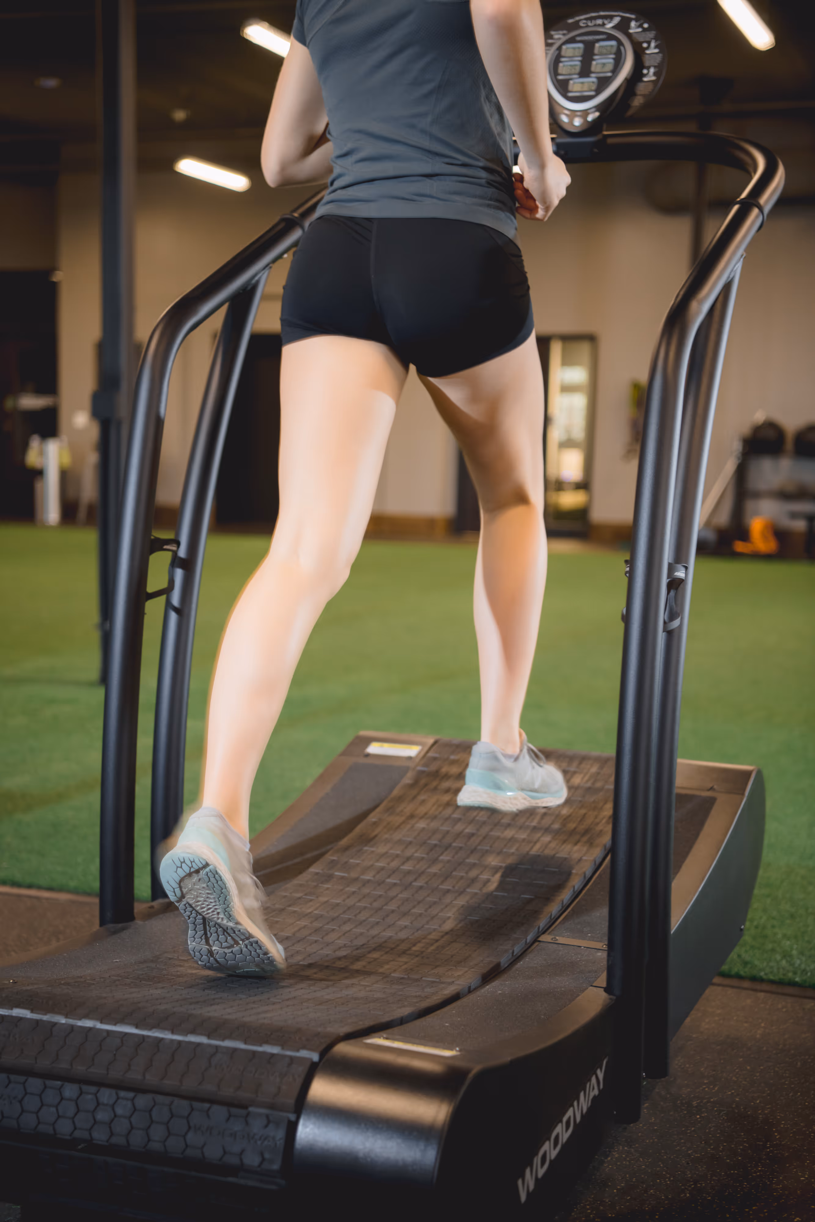
Proper bracing is crucial not only for lifting heavy weights in the gym, but also for lifting everyday objects, such as groceries, safely and effectively. Whether you’re performing squats, deadlifts, or picking up your nephew, mastering bracing techniques can enhance your performance and reduce the risk of injury.
Here is our guide to effectively engage your core:
- Begin by taking a deep breath. Imagine filling your abdomen with air.
- Contract your abdominal muscles, lower back, diaphragm, and pelvic floor.
- You don’t NOT want to push out your stomach and rib cage, but rather think of drawing everything in like a tight corset.
- This creates a feeling of tightness and tension in your core.
Remember, bracing is more than just holding your breath; it’s about creating stability and transmitting force effectively.
Preventing Diastasis Recti After Pregnancy
Diastisis recti during pregnancy is very common and the body’s natural way for making room for the growing baby. As the uterus expands during pregnancy, the abdominal muscles stretch and the linea alba separates. After childbirth, the linea alba can heal and come back together, but sometimes it doesn’t completely close. Learning how to properly brace your core and create leverage when lifting and carrying can be both helpful in prevention of diastasis recti, as well as promoting healing postpartum.
Treating Diastasis Recti on Your Own
Training with and through diastasis recti revolves around making that core area stronger and thus more functional for you. It is safe and effective to use and train your core while dealing with diastasis rectus. Treating diastasis should include recruiting deep core muscles in conjunction with those “6 pack abs” and more superficial core muscles.
If you are unsure how to recruit your deep core muscles, what it feels like, or points of performance with exercises/positions seeking help from a qualified physical therapist can help guide you in the process and take out any guess work while giving you the confidence that you are moving in the right direction to achieve your goals.
Seeking Professional Help for Diastasis Recti
If you suspect you have diastasis recti, it’s important to consult a healthcare professional. Again, diastasis recti is very common as it is a way our body makes room for the growing fetus. At a minimum, it’s important to have your doctor assess the severity of your diastasis recti, and then be guided through recommended lifestyle and exercise tips. Most doctors will check a new mom’s abdomen for diastasis recti at their 6-week postpartum check up, if not also during the final stages of pregnancy. Most women don't notice their diastasis until after giving birth.
If you experience bulging and discomfort for several months postpartum, it is recommended to seek a professional evaluation from your doctor or a qualified physical therapist. Just like with your recovery from surgery, giving birth or an injury, it’s important to have guidance of appropriate exercises, lifestyle modifications, and to be able to monitor your progress.
Surgical Interventions
Surgical interventions may be considered for diastasis recti when conservative measures fail to reduce the widening of the linea alba, and when discomfort is severe. A rectus plication, abdominoplasty, or tummy tuck, aims to reconstruct the linea alba and restore its anatomy by decreasing the width. The preoperative assessment typically involves plastic surgeons often requesting MRI scans for patients scheduled for abdominoplasty (tummy tuck) to repair diastasis recti.
The surgery involves tightening abdominal muscles to bring them closer together and decrease the width between. Surgery can be performed through open or laparoscopic surgery. Hernia repair surgery is another option that allows the ability to address both diastasis and any concurrent abdominal hernias. MRI helps evaluate the extent of separation, identify any associated hernias, and guide surgical planning.
Recovery among patients can vary. Swelling can typically take 6 weeks to subside. Recovery may also include 3-4 weeks of wear long stockings to prevent blood clots. Patients can typically return to office work and more sedentary demands 1-2 weeks after, however heavy lifting is often avoided for 2-4 weeks but can vary based on the patient, the surgeon and specific cases.
When Physical Therapy is Right for Diastasis Recti
Seeing a physical therapist for diastasis recti is beneficial because they can provide personalized guidance and treatment tailored to your specific needs. They can assess the severity of your condition and develop a customized exercise program to strengthen the core muscles, improve posture, and reduce strain on the abdominal muscles. Additionally, a physical therapist can teach you proper body mechanics and techniques for everyday activities to prevent further aggravation of the condition. They can also monitor your progress over time and make adjustments to your treatment plan as needed. Overall, working with a physical therapist can optimize your recovery and help you achieve better outcomes.